Liberia #16
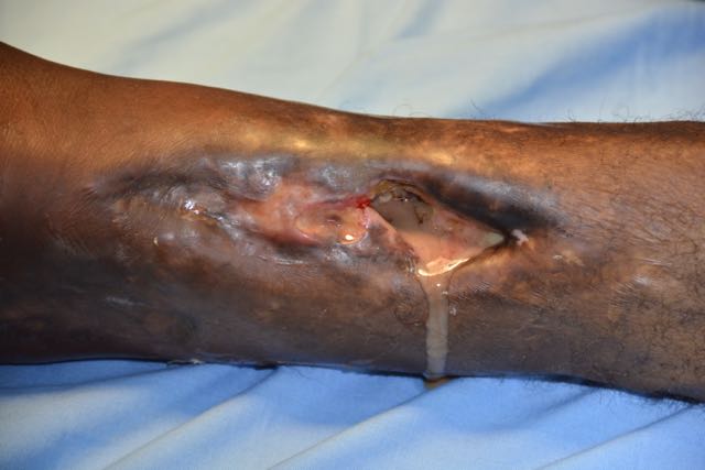
He lays on the operating room table moaning. He’s about 60 and covered in a hospital gown. Below his right knee a hole can be seen that is about an inch and a half wide and an inch deep and is a hole about the same depth. The hole is in his tibia. A terrible odor bathes the room as pus flows out of his bone. The ankle below is swollen to three times the normal size and feels soft and fluctuant. I put a needle in it last evening and got pus out. He told me that his leg has had the hole in it 10 years, and drains small amounts only. About a week ago his ankle became swollen and a couple days ago pus started draining from the hole. A septic ankle and chronic ostomyelitis are my diagnoses. I decide to use cautrey on this case, even though I suspect sterility of the device is suspect. Besides, how much more infected can I make a joint with pus in it? A spinal is attempted by the anestatist student, I seriously question sterility in all places, her teacher doesn’t really use sterile technique! I’ve seen him, dawn his sterile gloves then grab a bottle of rubbing alcohol to put on some gauze. (the outside of the bottle is NOT sterile). Everyone will get a longer course of antibiotics. After multiple bloody attempts, the anesthetist dawns his gloves and helps, getting it in. they patient lays back again. I insert a foley, STERILLY! Then I scrub, and dawn my apparel. I use the cautrey to make a cut vertically over the lateral ankle. Things look fine till I get down to the joint new level of stink, on the already pungent room. I can pass my finger from front to back along the side of the joint. So I open in the back as well. I flush in a liter or two of saline until it is coming out clear. We place a plastic flexible (penrose) drain and suture it in place. In the hole, mid-leg, I see dead bone, and pull out chunks. I bet there is more, but will stop at this for tonight. I suspect a sequestrium (retained dead bone fragment) but would like an xray before progressing. We don’t have one, so when he is a little better, will send him to another hospital for x-rays. (the referral hospital that is sending patients to us, JFK) His leg is wrapped with lots of gauze and then an ace bandage. He goes back to the floor when he is off oxygen and has stable vital signs. He is the last surgery of my day.
I began with rounds, right after the morning devotion at the hospital. I saw half of the 26 patients. Then I had to wait a bit till the OR team was ready to start. I did a small (by African standards) inguinal hernia with a bulge out all the time about 2 by 3 inches in size. I do a standard repair using mesh that has been sterilized in the autoclave. Then next one is similar, with two hernias that are larger.
“Doc, cam see emergency!” What’s wrong with them? “Pane to much de belly” As I follow Ruth downstairs to the cot in the room we call Emergency, I’m thinking of the different causes here. Appendicitis, typhoid perforation, ulcer perf, cancer perf, the list is much longer. A 14 year old boy is writhing around on the cot. I put a glove on and touch is abdomen. It’s rock hard and when I push down it causes pain, when I let up my hand quickly, he hollers in pain. Peritonitis! He will be the next surgery. They place an IV there and start Ampicillin and Chloramphenacol. I suspect appendicitis, but have been fooled by typhoid before, so I make a midline incision in the OR. Before the incision, we pray for him as is customary. Right under the skin is the fascia (the strength layer). As I open the peritoneum, pus flows up and drains down either side of his still abdomen. The amount we catch and goes to the suction canister is 250ml (half pint). Eventually I discover a necrotic appendix. I pull it up and tie off the base a couple times. I wash out the abdomen with many liters of saline, till clear. Then close up three layers. He should do much better now!